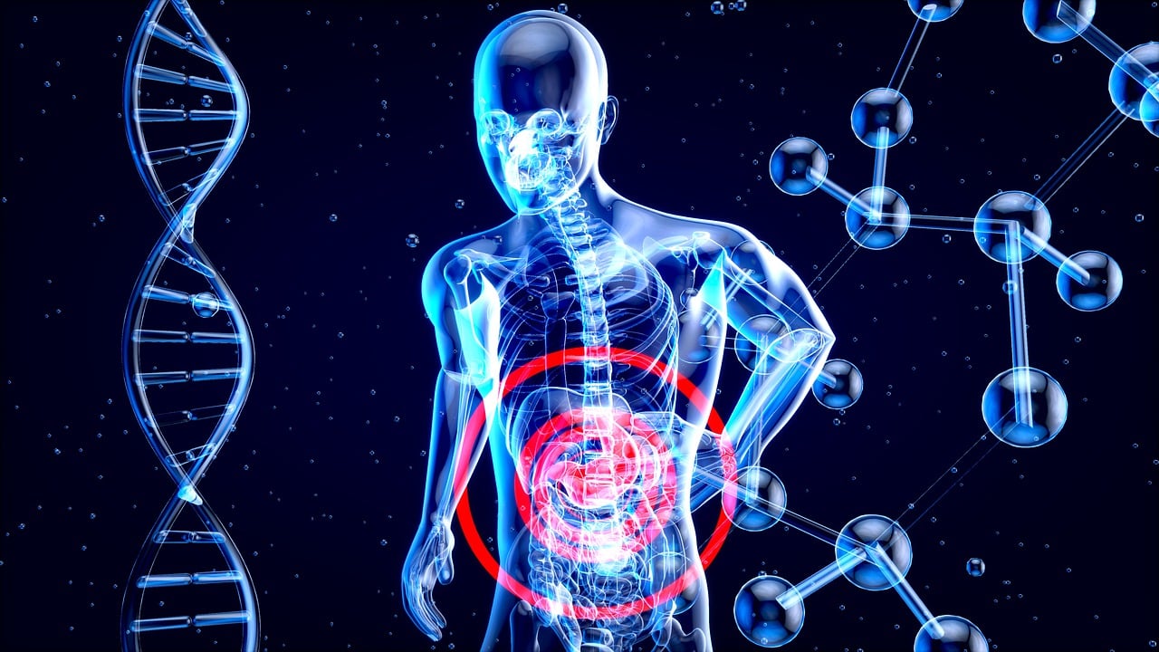
An X-ray is a medical imaging technology that uses high-energy electromagnetic radiation to create images of the inside of the body. The process involves passing X-rays through the body and onto a detector on the other side, which produces an image based on the X-rays that are absorbed or scattered by the body’s tissues and bones.
X-rays are often used to help diagnose bone fractures, infections, tumors, and other medical conditions. They are also used in dentistry to detect tooth decay and in some security screening systems to detect hidden objects.
X-rays are a form of ionizing radiation, which means they can be potentially harmful to living tissue if exposure is too high or frequent. However, the amount of radiation used in medical X-rays is typically very small and considered safe for most people.
Radiography
Radiography is a type of medical imaging that uses X-rays to produce images of the inside of the body. It is the most commonly used type of X-ray imaging and is used to diagnose a wide range of medical conditions.
During a radiography procedure, the patient is positioned between an X-ray machine and a detector, which captures the X-rays that pass through the body. The X-rays are absorbed or scattered differently by the various tissues in the body, and this creates an image on the detector that shows the internal structures of the body.
Radiography can be used to diagnose a variety of conditions, such as broken bones, lung infections, and digestive system problems. It is a non-invasive and relatively quick procedure that is widely available in hospitals and clinics.
However, as with any medical imaging technique that uses ionizing radiation, there is a small risk of radiation exposure with radiography. The amount of radiation used in a typical radiography procedure is generally considered safe for most people, but repeated exposure can increase the risk of developing cancer over time. Therefore, doctors and technicians take steps to minimize radiation exposure during the procedure, such as using lead aprons to shield the body from unnecessary radiation.
Computed tomography (CT)
Computed tomography (CT), also known as computed axial tomography (CAT), is a type of medical imaging technology that uses X-rays and computer processing to create detailed, cross-sectional images of the body.
During a CT scan, the patient lies on a table that moves through a doughnut-shaped machine that houses an X-ray tube and detectors. The X-ray tube rotates around the patient, emitting a series of narrow X-ray beams as it goes. The detectors measure the amount of X-rays that pass through the body at different angles, and a computer processes this information to create a series of 2D images (slices) of the body.
These images can be combined to create a 3D image of the body, which can be used to diagnose and monitor a wide range of medical conditions, including injuries, infections, and cancer. CT scans can be used to image almost any part of the body, including the head, chest, abdomen, and extremities.
Because CT scans involve exposure to ionizing radiation, there is a small risk of harm to the body’s cells. However, the benefits of the test often outweigh the risks, especially in cases where the information obtained can help guide medical treatment.
Fluoroscopy
Fluoroscopy is a type of medical imaging that uses X-rays to produce real-time images of the body. It involves passing a continuous beam of X-rays through the body and capturing the images on a video screen, similar to a continuous X-ray movie.
Fluoroscopy is often used during medical procedures, such as catheterization, to guide the placement of medical devices and to monitor the progress of the procedure in real-time. It can also be used to diagnose certain medical conditions, such as digestive problems or abnormalities in the urinary system.
During a fluoroscopy procedure, the patient lies on a table and the X-ray machine is positioned above or below the area being imaged. A contrast agent, such as barium or iodine, may be used to help highlight the area of interest and make it easier to see on the video screen.
While fluoroscopy is generally considered safe, it does involve exposure to ionizing radiation, which can be harmful in high doses. For this reason, the amount of radiation used during a fluoroscopy procedure is carefully controlled and kept as low as possible.
Mammography
Mammography is a type of medical imaging that uses X-rays to create images of the breast tissue. It is primarily used for breast cancer screening and diagnosis, as it can detect early signs of breast cancer before any physical symptoms are present.
During a mammogram, the breast is compressed between two plates and an X-ray is taken. This process may be slightly uncomfortable, but it only takes a few seconds to complete. The images produced by mammography can show abnormal lumps or masses, calcifications (tiny mineral deposits in the breast tissue), and other changes that may indicate the presence of breast cancer.
Mammography is recommended for women starting at age 40 (or earlier for women with a family history of breast cancer), and it is an important tool for early detection and treatment of breast cancer. However, like all medical procedures, mammography does have some risks, such as exposure to ionizing radiation, which can increase the risk of cancer over time. The benefits of mammography generally outweigh the risks, but women should discuss the risks and benefits with their healthcare provider before deciding whether to undergo screening.
Dental X-rays
Dental X-rays, also known as dental radiographs, are a type of X-ray imaging used to capture images of the teeth, gums, and jaws. These images can help dentists diagnose dental problems such as tooth decay, gum disease, and impacted teeth.
Dental X-rays use a small amount of ionizing radiation to create the images. The level of radiation exposure from a dental X-ray is generally considered to be very low, and the benefits of the images in diagnosing and treating dental problems usually outweigh any potential risks.
There are two main types of dental X-rays: intraoral and extraoral.
- Intraoral X-rays: These are the most common type of dental X-rays, and they involve placing a small sensor inside the mouth to capture images of individual teeth. There are several types of intraoral X-rays, including:
Bitewing X-rays: These are used to capture images of the back teeth (molars and premolars) and can help detect tooth decay and gum disease.
Periapical X-rays: These are used to capture images of individual teeth from the crown to the tip of the root and can help diagnose dental problems such as abscesses and root fractures.
- Extraoral X-rays: These X-rays capture images of the jaw and skull and are used to evaluate the position and development of the teeth, as well as to diagnose other conditions such as temporomandibular joint disorder (TMJ) and sleep apnea. There are several types of extraoral X-rays, including:
Panoramic X-rays: These capture a wide-angle view of the entire mouth, including all of the teeth, jaws, and other structures.
Cone-beam computed tomography (CBCT) scans: These are a type of 3D X-ray that can provide detailed images of the teeth, jaws, and other structures. They are often used for orthodontic treatment planning and to evaluate complex dental problems.


